
Scaphoid bone fracture
Browse 200+ scaphoid fracture stock photos and images available, or start a new search to explore more stock photos and images.
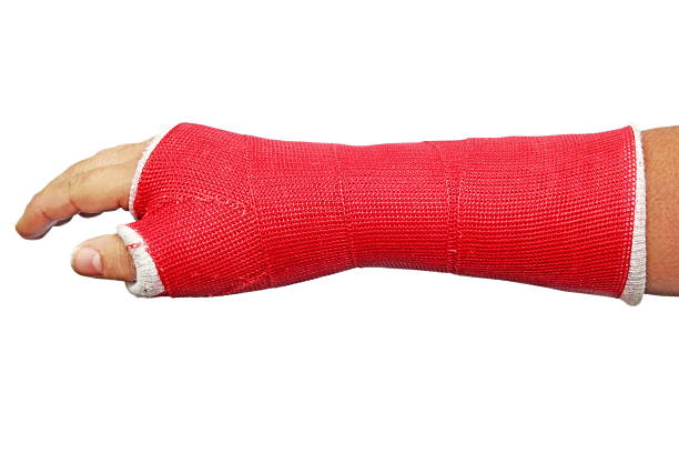
Orthopedic cast as treatment for scaphoid fracture

X-ray image of broken hand (Anteroposterior (AP) and oblique views), showing the 5th of metacarpal bone fractrue.

CT SCAN 3D of wrist,Lateral and AP position.

X-ray image of Left wrist joint AP view for showing fracture of radius bone.

A surgeon doctor examines a patient s palm for a fracture or crack in a bone. Scaphoid fracture, medical

Scaphoid bone fracture medical vector illustration on white background eps 10

Three month later navicular bone fracture. Pseudoarthrosis complication. No fracture healing.

X-ray image of wrist joint Ap view for diagnostic rheumatoid arthritis . X-ray image of wrist joint Ap view for diagnostic rheumatoid arthritis .

X-ray wrist PA view under slab with distal radius fracture ,Medical image concept.

Polydactyly (hyperdactyly) refers to the situation where there are more than the usual number of digits (five) in a hand or foot

X-ray Left wrist joint AP,LATERAL Fracture with displacement distal end left radius.Soft tissue swelling.
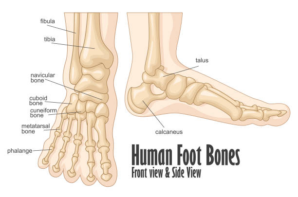
Vector Illustration Of Human foot bones front and side view anatomy

bones of the foot and ankle joint medical vector illustration isolated on white background eps 10

Abdominal aneurysm 3d medical vector illustration isolated on white background eps 10 with description
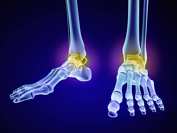
Skeletal foot - injuryd talus bone. Xray view. Medically accurate 3D illustration

Illustration of the skeletal foot

X-ray image of Right wrist joint Ap and Lateral view for diagnosis rheumatoid arthritis .

X-ray image of broken hand, PA and lateral view. Show metacarpal fracture.

X-ray image of Right wrist joint Ap view for diagnosis rheumatoid arthritis .

3D Illustration of Human Skeleton System Tibia and Fibula Anatomy Anterior View

X-ray image of the patient’s hands after bilateral amputation of fingers, anteroposterior (AP) view, diabetic patient

Skeletal foot - injuryd talus bone. Xray view. Medically accurate 3D illustration

Vector Illustration Of Human foot bones front and side view anatomy

Illustration of the Foot

Polydactyly (hyperdactyly) refers to the situation where there are more than the usual number of digits (five) in a hand or foot

X-ray Left wrist joint Fracture with displacement distal end left radius.Soft tissue swelling.

A photograph of the fingers and hand is applied to the patient's hand. The radiologist examines the x-ray. X-ray of a woman's wrist.

X-ray image of wist joint, PA and lateral view. Showing distal radius fracture.

Hand scar and affected skin due to medical operation after a bone fracture.

X-ray image of wrist joint, PA view. Showing radius fracture.

Digital wrist joint x-ray image with wooden splint, AP view, shows radius fracture.

X-ray image of Left wrist joint AP and Lateral view for showing fracture of radius bone.

X-ray image of Hand, oblique view. Shows proximal of middle finger fracture

anatomy, horizontal, human body part, human joint, data, medical x-ray, wrist, bone fracture, human bone, human skeleton, medical scan, no people, science, close-up, human arm, human limb, joint - body part, radius bone, ulna, biology, hospital, indoors, patient, bone, orthopedics, tomography, technology, human finger, navicular bone of hand, physical injury, bending, doctor, finger, illness, pain, pointing, problems, radiation, shoulder, surgery, crash, medical exam, osteoporosis, symptom, arm bone, medical condition, mri scan, limb - body part, metacarpal, inflammation

X-ray of the foot broken calcaneal/Heel with copy space.

X-ray Left wrist joint Fracture with displacement distal end left radius.Soft tissue swelling.

3D Isometric Flat Vector Conceptual Illustration of Distal Radius Fracture, Labeled Educational Diagram


