
Shoulder arthroscopy procedure. Rotator cuff tears or shoulder impingement. Minimally invasive surgery. Arthroscopic joint treatment, pain and inflammation in human body x ray flat vector illustration
Browse 310+ shoulder arthroscopy stock photos and images available, or search for arthroscopic surgery to find more great stock photos and pictures.
Shoulder arthroscopy procedure. Rotator cuff tears or shoulder impingement. Minimally invasive surgery. Arthroscopic joint treatment, pain and inflammation in human body x ray flat vector illustration
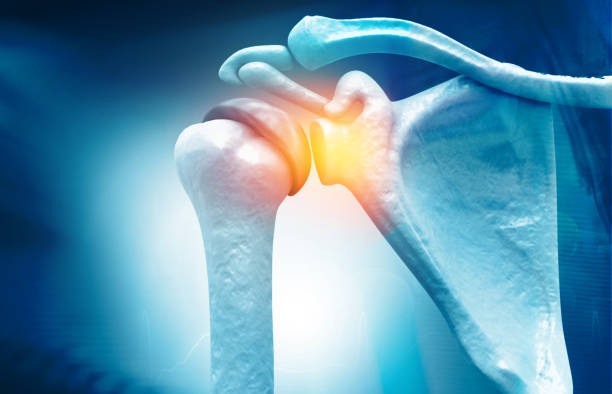
Anatomy of Shoulder , rotator cuff tear, Shoulder pain. 3d illustration

Man with painful shoulder joint with blue background - x-ray 3d illustration

Woman with painful shoulder joint with blue background - x-ray 3d illustration

Male shoulder with scars after shoulder Arthroscope surgery. Chronic dislocation of the long biceps tendon after surgery. Reconstruction of the cranial third of the SSC tendon corkscrew anchor. Wound

Shoulder arthroscopy procedure. Rotator cuff tears or shoulder joint replacement. Minimally invasive surgery. Ligaments treatment, tendonitis pain or arthritis inflammation medical vector illustration

Arthroscopic view - from the bursal side - of the torn supraspinatus tendon near the insertion site on the greater tubercle of the right shoulder. A soft tissue resector (shaver) has been introduced for debridement before the (suture anchor) repair (to enhance the healing).

Shoulder arthroscopy procedure. Subacromial decompression, bursitis or shoulder joint replacement. Minimally invasive surgery. Pain or arthritis inflammation in human body medical vector illustration

X Ray Illustration of Painful Shoulder Joint - Medical 3D Illustration

Shoulder arthroscopy procedure. Rotator cuff tears or shoulder joint replacement. Minimally invasive surgery. Ligaments treatment, tendonitis pain or arthritis inflammation medical vector illustration

Shoulder arthroscopy procedure. Subacromial decompression, bursitis or shoulder joint replacement. Minimally invasive surgery. Pain or arthritis inflammation in human body medical vector illustration

Reconstruction of the cranial third of the SSC tendon corkscrew anchor. Wound

X-ray 3D Illustration of Pain in Shoulder Joint with blue Background - 3D Illustration

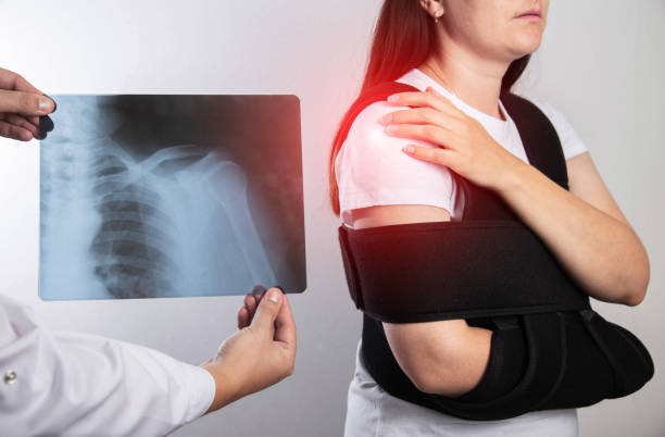
A doctor surgeon examines a girl's shoulder after surgery. Bandage on the shoulder joint during rehabilitation after surgery on the shoulder joint, tear of the shoulder cuff.

Reconstruction of the cranial third of the SSC tendon corkscrew anchor. Wound

A girl in a white T-shirt is holding her shoulder joint and is in severe pain. Concept of habitual shoulder dislocation. Improper rehabilitation, surgery to treat a dislocated shoulder.

Reconstruction of the cranial third of the SSC tendon corkscrew anchor. Wound

Shoulder arthroscopy procedure. Rotator cuff tears or shoulder impingement. Minimally invasive surgery. Arthroscopic joint treatment, pain and inflammation in human body x ray flat vector illustration

Doctor surgeon holds a scalpel against the background of the shoulder of the girl concept of surgery on the shoulder joint and the replacement of an artificial joint, arthroplasty, capsular ligament apparatus

Shoulder arthroscopy procedure. Rotator cuff tears, shoulder muscles problem. Minimally invasive surgery. Arthroscopic joint and ligaments treatment, pain or inflammation in the human body flat vector

Arthroscopic view (from the posterior portal) of a left human shoulder with a partial thickness rupture of the supraspinatus tendon (PASTA lesion of the rotator cuff) at the articular side of the footprint of the tuberculum majus. The long biceps tendon is seen in the background to the right. The humeral head is seen in the lower part of the image. Smaller PASTA lesions are treated by debridement only. Larger lesions should be repaired.

Arthroscopic view (from posterior portal) of a left shoulder during repair of a SLAP II lesion (avulsion of the superior part of labrum including biceps anchor). A suture grasper (or manipulator) is being used to retrieve the two suture ends (one on each side of the biceps anchor) of a suture anchor used for repairing the labral tear.

A girl holds an X-ray of a dislocation and fracture of the humerus in a bandage on the shoulder joint after a surgical operation.

Reconstruction of the cranial third of the SSC tendon corkscrew anchor. Wound

Arthroscopic view, from a lateral portal, of a large U-shaped tear of the rotator cuff including both the supraspinatus and infraspinatus tendons. The first of a series of side-to-side sutures has been placed (with a special arthroscopic sewing device). The orthopaedic surgeon is securing a sliding knot with a knot pusher causing the rupture ends of the tendons to approach each other. The arthroscope is placed in the subacromial space, but due to the massive tear, the intra-articular components are revealed including the humeral head, glenoid, labrum and the long head of the biceps brachii (LHB) tendon. The image was captured with a 4mm 30 degrees arthroscope. A copy of the same image with explaining text (labels) added onto the image, to aid a designer with limited medical knowledge, has been included in the same series.

Arthroscopic view (from posterior portal) of a left shoulder suffering from adhesive capsulitis (or frozen shoulder). The rotator interval (between the long head of the biceps and subscapularis tendon) if filled with inflamed synovium, adhesions and scar tissue. The humeral head is in the lower-left corner.

Arthroscopic view of repair of a torn rotator cuff (supraspinatus tendon) of the right (human) shoulder. The repair will be finalised by fixing the torn tendon to the greater tubercle (tuberculum majus) of the humerus by a suture anchor. The image shows the placement of the suture anchor. The image was captured with a 4mm 30 degrees arthroscope introduced through a lateral portal by an orthopaedic surgeon (who is also the photographer).

Woman with painful shoulder joint with blue background - x-ray 3d illustration

Reconstruction of the cranial third of the SSC tendon corkscrew anchor. Wound

Woman with painful shoulder joint with blue background - x-ray 3d illustration

Arthroscopic view - from the articular side - of the torn supraspinatus tendon and part of the anterior part of the infraspinatus (of the rotator cuff) near the insertion site on the greater tubercle of the right shoulder. The U-formed defect in the tendon allows the surgeon to look directly through the joint into the subacromial space. After debridement of the footprint (of the supraspinatus tendon), a cuff repair was performed. The long head of the biceps muscle is seen to the left in the image.

Arthroscopic view, from a lateral portal, of a large U-shaped tear of the rotator cuff including both the supraspinatus and infraspinatus tendons. The first of a series of side-to-side sutures has been placed (with a special arthroscopic sewing device). The orthopaedic surgeon is securing a sliding knot with a knot pusher causing the rupture ends of the tendons to approach each other. The arthroscope is placed in the subacromial space, but due to the massive tear, the intra-articular components are revealed including the humeral head, glenoid, labrum and the long head of the biceps brachii (LHB) tendon. The image was captured with a 4mm 30 degrees arthroscope. Text (labels) has been added onto the image to facilitate the use by a designer that may not be a medical scholar. A copy of the same image without the text has been included in the same series.

Arthroscopic view (from posterior portal) of a right shoulder suffering from adhesive capsulitis (or frozen shoulder). An anterior capsulotomy - marked with dotted line - is being performed by a meniscal (upbiter) punch.

Arthroscopic view (from a posterior portal) of a sliding knot being tightened (over the biceps anchor of the right shoulder) with help of a knot pusher (inside a clear plastic cannula) as part of a repair of a SLAP lesion - a tear of the superior part of the labrum including the biceps anchor.

Arthroscopic view (from posterior portal) of a right shoulder suffering from adhesive capsulitis (or frozen shoulder) - in an early stage with synovitis and pain.

Shoulder arthroscopy procedure. Rotator cuff tears or shoulder impingement. Minimally invasive surgery. Arthroscopic joint treatment, pain and inflammation in human body x ray flat vector illustration

Arthroscopic (keyhole) view (from the posterior portal) of a right shoulder during repair of a Bankart lesion (avulsion of the anterior-inferior part of the labrum from the glenoid - resulting in instability and dislocations) with three suture anchors.

Arthroscopic view, from a lateral portal, of a large U-shaped tear of the rotator cuff including both the supraspinatus and infraspinatus tendons. The arthroscope is placed in the subacromial space, but due to the massive tear, the intra-articular components are revealed including the humeral head, glenoid, labrum and the long head of the biceps brachii (LHB) tendon. The image was captured with a 4mm 30 degrees arthroscope. A copy of the same image with explaining text (labels) added onto the image, to aid a designer with limited medical knowledge, has been included in the same series.
The doctor holds in his hand a medical x-ray of a dislocated humerus and a fractured collarbone against the background of a girl patient whose shoulder hurts. Fixing bandage for the shoulder joint. Tramatology and orthopedics, close-up

Arthroscopic view (from posterior portal) of a right shoulder suffering from adhesive capsulitis (or frozen shoulder). A soft tissue resector (or shaver) is being introduced in the rotator interval (between the long head of the biceps and subscapularis tendon) to remove inflamed synovium and scar tissue. The humeral head is in the lower-right corner.

Arthroscopic view (from posterior portal) of a left shoulder during repair of a SLAP II lesion (avulsion of the superior part of labrum including biceps anchor). A suture anchor has been inserted into a drill hole in glenoid. A sharp suture retrieving instrument piercing the base of the avulsed labrum has grasped one of the suture ends and is about to retract and pass the suture reversely.

Arthroscopic view of a titanium suture screw anchor about to be inserted into the debrided foot print of the torn supraspinatus tendon on the greater tubercle of the humerus of the right shoulder. The device consists of a titanium screw with an eyelet with two strong sutures for suturing the torn rotator cuff tendon end (supraspinatus in the current case) back onto the bony surface. The image was captured with a 4mm 30 degrees arthroscope. Text (labels) has been added onto the image to facilitate the use by a designer that may not be a medical scholar. A copy of the same image without the text has been included in the same series.

Arthroscopic view (from a lateral portal) of the subacromial space of a right shoulder. A burr (acrominizer) - through a posterior portal - is used for acromioplasty (revealing spongious bone of the underside of the acromion).

Shoulder arthroscopy procedure. Rotator cuff tears or shoulder impingement. Minimally invasive surgery. Arthroscopic joint treatment, pain and inflammation in human body x ray flat vector illustration

Shoulder arthroscopy procedure. Subacromial decompression, bursitis or shoulder joint replacement. Minimally invasive surgery. Pain or arthritis inflammation in human body medical vector illustration

Shoulder dislocation. humerus bone trauma, Sports injuries, or Weak shoulder muscles. Human arm anatomy. Bones and joint of the Shoulder, and hand. Vector illustration

Arthroscopic view (from a lateral portal) of the subacromial space of the right shoulder with a U-shaped rupture of the supraspinatus tendon - marked with a dotted line. A needle (N) with attached (blue) suture has penetrated both parts of the tear and is about to be retrieved with a needle holder (NH). A tissue grasper (TG) is seen in the background. The (long head of the) biceps tendon is thickened and frayed.

Arthroscopic view (from posterior portal) of a right shoulder suffering from adhesive capsulitis (or frozen shoulder). Inflamed synovium and scar tissue have already been removed from the rotator interval. Now the Middle Glenohumeral Ligament is being released (cut) by a meniscal (upbiter) punch.

A simplified 3D rendering of a shoulder joint with basic colors and textures to highlight areas of calcific tendonitis.

Shoulder arthroscopy procedure. Subacromial decompression, bursitis or shoulder joint replacement. Minimally invasive surgery. Pain or arthritis inflammation in human body medical vector illustration

A girl has pain in her shoulder due to a pinched nerve in the cervical spine, osteochondrosis of the spine. Referred pain

Orthopedic Surgeon: Woman's Healing Journey After Shoulder Surgery In Hospital

Arthroscopic view of repair of a torn rotator cuff (supraspinatus tendon) of the right (human) shoulder. A special suture passing device (SPD) “sewing machine” used for rotator cuff repair in the shoulder. A needle loaded with a suture end penetrates the supraspinatus tendon. The torn tendon gap is closed by side-by-side sutures. The repair will be finalised by fixing the torn tendon to the greater tubercle (tuberculum majus) of the humerus by a suture screw anchor made of titanium. The image was captured with a 4mm 30 degrees arthroscope introduced through a lateral portal by an orthopaedic surgeon (who is also the photographer). The image is part of a series.

A girl in white with a fixing black bandage on her arm after an injury, a fracture of the radius and a sprain. Arm support bandage

Shoulder arthroscopy procedure. Rotator cuff tears, shoulder muscles problem. Minimally invasive surgery. Arthroscopic joint and ligaments treatment, pain or inflammation in the human body flat vector

Arthroscopic intra-articular view from a posterior portal of a moderately large U-shaped tear of the rotator cuff (supraspinatus tendon) of the right (human) shoulder. A motorized 5.5mm soft tissue resector (shaver or synovator) has been introduced through a lateral portal and is being used to debride (remove or clean up) the footprint of the supraspinatus tendon (at the greater tubercle or tuberculum majus humeri) to facilitate healing after it has been repaired (fastened to the bone with a suture anchor). The image was captured with a 4mm 30 degrees arthroscope. Text (labels) has been added onto the image to facilitate the use by a designer that may not be a medical scholar. A copy of the same image without the text has been included in the same series.


